What are the eye exams?

What are the eye exams?
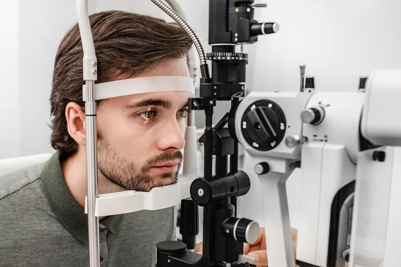
Slit lamp: A device with a strong light source that help to examine the anterior parts of the eye, starting from the eyelids, the cornea, the anterior chamber of the eye, the pupil and the lens.
Tonometry: A device to measure the eye pressure to follow up on its measurement, especially for patients with glaucoma.
Sound waves:
-It is used when there is an obstacle that prevents seeing what is inside the eye, such as corneal opacity, cataracts, or bleeding in the vitreous body of the eye.
– It is used to measure the extent of retinal detachment and the dimensions of tumors inside the eye, and to measure the eye and the thickness of the eye muscles.
-Visual strength check.
– Important to assess the patient’s ability to see things at specific distances, and it is important for measuring glasses and contact lenses.
Gonioscopy:
-It is used to examine the corners of the anterior chamber of the eye. This part is responsible for draining the fluid in the anterior chamber.
-A basic examination to assess cases of glaucoma uses a special type of lenses to perform this examination.
Ophthalmoscopy:
–It used to examine the retina is usually a wide pupil to help see a larger field than the retina. There are two types, direct and indirect.
-Direct helps the doctor see the human nerve and the central part of the retina, while the peripheral parts of the retina can be seen well using indirectly.
Phoropter: A device with a set of lenses placed in front of the patient’s eye and the patient looks through it at the marking board and through which the power of the lens can be changed to reach for the right lens for eyeglasses corneal imaging.
Pentacam: Visualizing the details of the corneal surface the computer makes a color image of the corneal surface in terms of astigmatism and corneal diseases.
Keratometry: The corneal imaging device maps more accurate details when it is difficult to obtain accurate data from the device For the surface of the cornea, it is important to know the details of the cornea to make accurate measurements for vision correction operations and to calculate the Steepest & flattest.
– used to measure Lens strength Eye fundus pictures.
-It is required to record the state of the eye nerve, visual center, retina and vitreous body. A special type of camera is used to photograph the progression of the disease, such as:
– Atrophy of the visual center or the effect of diabetes on the retina X-ray of the retina of the eye.
-It is used to evaluate the effect of various symptoms on the retina of the eye. This examination requires injecting a dye substance through the patient’s arm and within seconds it takes effect, the dye is in the blood vessels until it reaches the blood vessels inside the eye. Pictures are taken to record any infiltration of the dye inside the eye, which helps the doctor on diagnosing the condition.
O.C.T:
-CT scan of the optic nerve and retina, which takes pictures of the retina with cross-sectional images, which takes less than 10 minutes, It can photograph any change, even a slight change, in the retina, which is of great benefit to glaucoma and retinal disease and visual center injuries amsler,A quick examination helps assess the state of the visual center, which is small squares in the middle of these The squares are a point and the patient is asked to look at the point and notice if there is any change in the squares, which is looking at the point Field of vision. It assesses the patient’s field of vision because it is one of the things that are affected by some diseases such as glaucoma, and it also assesses the neurological capacity of the retina and nerve, The eye and the brain, which requires the patient to focus on a specific point and press a certain button when he sees a flash of light.


