Al-Dokki BranchNasr City BranchBeni-Suef Branch
Blog

Blog list

Compellingly embrace empowered e-business after user friendly intellectual capital. Interactively actualize front-end processes with effective convergence. Synergistically deliver performance based methods of empowerment.

10.webp
30/Oct/2022

What is the squint?

It is a visual defect in the position of the two eyes, where each point in a different direction, one of the eyes may be straight (pointing forward) while deviating the other eye is inward or outward, first up or down.

The imbalance of the position of the eyes may be observed continuously, or it appears and disappears at other times, and this imbalance may be transmitted between the eyes and this condition usually appears in Children, and the percentage of its appearance in the hoofs in the United States, for example, is 4%. It also appears in advanced stages of age. Amblyopia appears in the same percentage in males. And females and may be genetically transmitted from fathers to sons, but there are exceptions to this rule.

How do the eyes work together?

In the case of a healthy person, the eyes point to the same point, and the brain combines the two images in the form of an image, One is three-dimensional, which gives the sense of depth, and when one of the eyes deviates, two images are sent When this condition appears in a young child, the brain learns to ignore the image from the brain The deviated eye sees only the image from the straight or better-seeing eye and as a result loses Baby’s sense of depth.

When strabismus appears in adults, they often have double vision because the brain is trained to receive image, from both eyes and cannot ignore the image from the deviated lazy eye.

Good vision is formed during early childhood and both eyes are in a straight position.

The brain accepts the image from the healthy eye and ignores the image from the weak, lazy eye, and this appears in about 50% of children for people with amblyopia.

Lazy eye can be treated by covering the healthy eye and improving vision in the weak eye. Usually, treatment succeeds if it is done. Diagnosis of lazy eye in the first years of life, but if treatment is delayed, lazy eye becomes a permanent condition. In the child, and as a rule, the earlier the diagnosis of lazy eye is, the better the vision will be children.

What is the reason of squint?

The sure cause of strabismus is not fully understood. We must know that there are six muscles that control the strabismus One eye movement and these muscles connect to the eye from the outside, and in both eyes there are two muscles that move the eye Right and left, while the other four muscles move the eye up and down and to any oblique angle The muscles in both eyes must work together in parallel to parallel the direction of the eyes and to determine the direction of vision in a point ,One and this allows the movement of the eyes in the same coordinated way The brain controls the movement of these muscles, so strabismus appears in some children who suffer from it of the following cases:

-Cerebral palsy.

-Mongolian.

-Hydrocephalus.

-Brain tumors.

-Strabismus can also result from cataract surgery or because of a severe eye injury.

What are the symptoms of strabismus?

The main symptom of strabismus is the presence of an eye that is not straight and in some cases, children with strabismus deviate one of their eyes in sunlight, Shining or tilting their heads in an attempt to use both eyes together.

How is amblyopia diagnosed?

Strabismus can be diagnosed through an eye examination, so children’s eyes are examined by an ophthalmologist before or at, The age of four, and in the event of previous cases of strabismus or lazy eye in the child’s family, it must be examined before the age of four the third.

How is amblyopia treated?

The main purpose of treatment is Maintaining strong eyesight Correction of the position of straight eyes Restoring the healthy vision of both eyes, and the ophthalmologist decides the method of treatment after a comprehensive eye examination, either by wearing Medical glasses or surgery to correct balance in the eye muscles or to remove cataracts, if present, and cover The healthy eye to treat lazy eye in the weak eye.

What are the types of alcohol?

Medial or internal strabismus:

It is a strabismus resulting from the deviation of the eye to the inside, and it is the common type of strabismus in children.

Children with amblyopia do not use their eyes together, and early surgery is usually necessary to adjust the position of the eyes

The doctor adjusts the tension on the muscles of one or both eyes, and this adjustment reduces the attraction of the eye inward and allows it to move more freely.

There is another way in which the doctor shortens the external muscle to allow the eye to move outward.

Adaptive lateral strabismus:

This type of strabismus is common among children with farsightedness at the age of two years or more. When a child is small or has farsightedness, His attempt to focus his gaze on objects close to the eye, which leads to the return of the eye to its normal position. Special eye drops and ointments help or Prismatic lenses to correct the position of the eye.

Lateral or extrinsic strabismus:

It is a strabismus resulting from the deviation of the eye outward and appears when the child strains his eyes in an attempt to focus his eyes on distant objects. Brutal strabismus appears, only from time to time, especially when he is stray, tired or sick, as parents notice that one of their child’s eyes deviates outward when facing a light Bright sun although glasses or prisms with eye exercises may help correct the position of the eye, surgery may be necessary.

How is strabismus surgery performed?

The ophthalmologist makes a small opening in the tissue surrounding the eye in order to reach the muscles surrounding it. The position of the eye muscles is corrected according to the direction of the eye deviation and strabismus correction surgery is performed in children under the influence of general anesthetic after surgery, the patient may need glasses or prisms, and in some cases, he may need another surgery to keep the eyes in the normal position.

For children with persistent squint, early surgery provides an excellent opportunity for the eyes to function normally, and the best for the child is this procedure

Surgery before school ageLike any other surgery, there may be some risks to the surgery, such as infection, bleeding or severe limping, which may lead to the loss of part of vision.

Strabismus surgery is a safe and effective procedure for treating eye deviation, but it does not dispense with the use of eyeglasses or the treatment of lazy eye caused by strabismus

What is new in the treatment of strabismus?

The American Food and Drug Administration has approved the limited use of a new drug called (Botox) as an alternative to strabismus surgery. This drug is injected into Deviated eye muscles, which leads to relaxation and tension of the opposite muscles, so the eye returns to its normal position, although the effect of the drug remains in the body for several only weeks, but in some cases it leads to permanent correction.


امراض-العين-الشبكية.webp
30/Oct/2022

What is the meaning perforation?

With age, the amount of fluid in the vitreous shrinks and begins to separate from the surface of the retina, which leads to a detachment in the retina.

What are the symptoms?

Blurred vision and poor visual acuity.

 What is meant by retinal artery occlusion?

The blockage of the central retinal artery causes vision loss after stopping the blood supply to the retina (tissue Nervousness of the eye) and that this phenomenon is rare.

Symptoms of retinal artery occlusion?

The symptoms of retinal artery occlusion are the feeling of a sudden and unaccompanied decrease in vision. Pain, which is represented by poor vision ranging from the ability to count the fingers to the sense of light only. There are sometimes predictive symptoms represented in short and expressive cases of loss of vision or seeing blackness, and there are cases, in which the injury is to one of the branches of the central artery, and thus the loss of vision is in part of the field of vision only.

Causes of retinal artery occlusion?

The most common cause of blockage is clots from the carotid artery, which necessitates examinations in vascular surgery.

There are many different factors and diseases that cause blockage, such as high blood pressure, diabetes, and blood clotting defects. Valve diseases Heart, inflammatory diseases in the blood vessels, autoimmune diseases. Tumors and fat emboli in injury and fractures in the pelvic area.

Retinal artery occlusion treatment?

Treatment will target three things:

 Removing the obstruction: reducing the pressure inside the eye

-Oxygen augmentation: suctioning a mixture of oxygen and carbon dioxide to enable oxygen to reach the retina from nearby tissues.

-Use of clot-dissolving agents.

-Determine the cause of the blockage.

-Attention to preventive treatment of paramount importance.

What is retinitis pigmentosa?

A group of diseases that often have genetic factors and cause eye atrophy, and the disease condition increases with disease from Night blindness leads to loss of the field of vision, and the disease can begin at any age, but most often it begins in adults.

What are the symptoms of retinitis pigmentosa?

-Night blindness in the early stages.

-Gradual loss of vision in advanced stages of life.

What are light flashes and dark spots?

Light flashes are rapid brightness of light in the field of vision intermittently dark spots occur, As a result of a disorder of the vitreous body.

What are the symptoms of light flashes and dark spots?

In the field of vision suddenly and the spots are sometimes mobile and it is possible that be accompanied by a gradual loss of vision.

What are the treatments ways?

In some cases, surgical treatment is not necessary and the patient can live with the condition, in advanced cases, the doctor advises surgical intervention to prevent any complications.


فحوصات-العين.webp
30/Oct/2022

What are the eye exams?

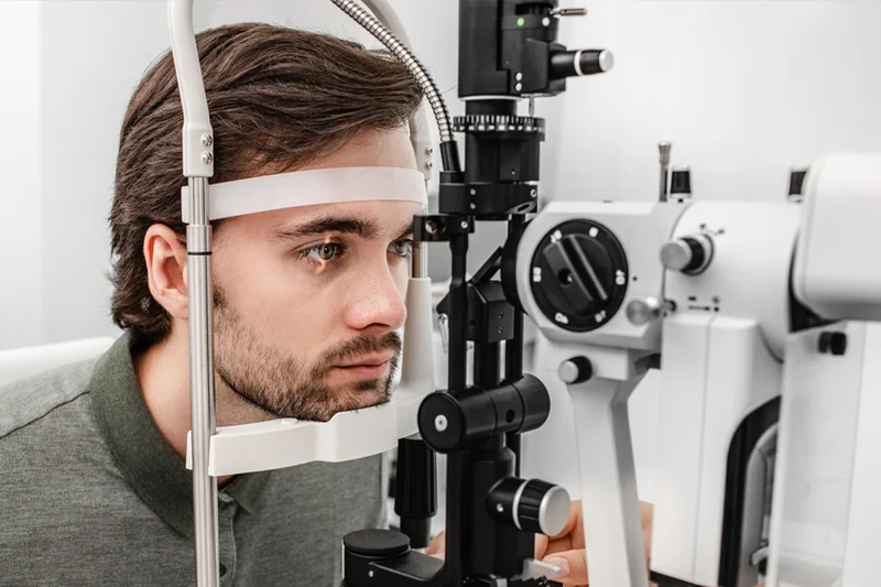
Slit lamp: A device with a strong light source that help to examine the anterior parts of the eye, starting from the eyelids, the cornea, the anterior chamber of the eye, the pupil and the lens.

Tonometry: A device to measure the eye pressure to follow up on its measurement, especially for patients with glaucoma.

Sound waves:

-It is used when there is an obstacle that prevents seeing what is inside the eye, such as corneal opacity, cataracts, or bleeding in the vitreous body of the eye.

– It is used to measure the extent of retinal detachment and the dimensions of tumors inside the eye, and to measure the eye and the thickness of the eye muscles.

-Visual strength check.

– Important to assess the patient’s ability to see things at specific distances, and it is important for measuring glasses and contact lenses.

Gonioscopy:

-It is used to examine the corners of the anterior chamber of the eye. This part is responsible for draining the fluid in the anterior chamber.

-A basic examination to assess cases of glaucoma uses a special type of lenses to perform this examination.

Ophthalmoscopy:

It used to examine the retina is usually a wide pupil to help see a larger field than the retina. There are two types, direct and indirect.

-Direct helps the doctor see the human nerve and the central part of the retina, while the peripheral parts of the retina can be seen well using indirectly.


Phoropter: A device with a set of lenses placed in front of the patient’s eye and the patient looks through it at the marking board and through which the power of the lens can be changed to reach for the right lens for eyeglasses corneal imaging.

Pentacam: Visualizing the details of the corneal surface the computer makes a color image of the corneal surface in terms of astigmatism and corneal diseases.

Keratometry: The corneal imaging device maps more accurate details when it is difficult to obtain accurate data from the device For the surface of the cornea, it is important to know the details of the cornea to make accurate measurements for vision correction operations and to calculate the Steepest & flattest.

– used to measure Lens strength Eye fundus pictures.

-It is required to record the state of the eye nerve, visual center, retina and vitreous body. A special type of camera is used to photograph the progression of the disease, such as:

– Atrophy of the visual center or the effect of diabetes on the retina X-ray of the retina of the eye.

-It is used to evaluate the effect of various symptoms on the retina of the eye. This examination requires injecting a dye substance through the patient’s arm and within seconds it takes effect, the dye is in the blood vessels until it reaches the blood vessels inside the eye. Pictures are taken to record any infiltration of the dye inside the eye, which helps the doctor on diagnosing the condition.

O.C.T:

-CT scan of the optic nerve and retina, which takes pictures of the retina with cross-sectional images, which takes less than 10 minutes, It can photograph any change, even a slight change, in the retina, which is of great benefit to glaucoma and retinal disease and visual center injuries amsler,A quick examination helps assess the state of the visual center, which is small squares in the middle of these The squares are a point and the patient is asked to look at the point and notice if there is any change in the squares, which is looking at the point Field of vision. It assesses the patient’s field of vision because it is one of the things that are affected by some diseases such as glaucoma, and it also assesses the neurological capacity of the retina and nerve, The eye and the brain, which requires the patient to focus on a specific point and press a certain button when he sees a flash of light.


ليزك.webp
30/Oct/2022

What is LASIK?

It is the modern technique for correcting vision using an intense light belt inside the cornea and not on the surface.

What are the advantages of this method in comparison to the old way?

The advantages are summarized in the accuracy of adjusting the measurements, the speed of corneal healing, the patient’s return to his activity within 24 hours, and the scarcity of any relapse in sight size.

What are the uses of the LASIK process?

LASIK can be used to correct the following vision defects

-Nearsightedness

-Farsightedness

-Astigmatism

The maximum correction depends on several factors such as corneal thickness, pupil size, and topography.

For the eye, and there are no contraindications inside the eye, such as iritis, cataracts, or glaucoma.

What is the age to do surgery?

The operation can be performed at any age, but according to the circumstances of each case, meaning that this operation can be performed for children when there is a big difference

In the size of the glasses between the two eyes, in this case, the operation can be performed for children when there is a large difference in the size of the glasses between the two eyes in this case.

The case can be performed for the weak eye if the child is unable to bear the medical glasses so as not to weaken the eye

It is possible in the case of the elderly, provided that there are no other eye diseases, as mentioned previously. As for the most appropriate age in general, it is 18 years and the reason for this is due to the increase the stability of the glasses size at this age and so that we do not have to repeat the LASIK operation again.


Is it possible to repeat the LASIK operation again?

Of course it is possible, after the process of re-doing the thickness and topography of the cornea.

What does the patient do before the operation?

 It is preferable not to wear soft contact lenses for at least two weeks before the operation, and hard lenses.

-4 weeks before the operation.

-It is possible to eat food before the operation, but light food, and it is preferable to go to the hospital or center with someone.

-Relatives or friends, and the doctor performs some accurate eye examinations directly before the operation.

What are the steps in the operating room and the time taken?

 The operation does not take more than ten minutes, and the operation is performed under the influence of local anesthesia (anesthetic drops), and the patient does not feel any pain during the operation.

-Then he feels a slight burning sensation for two to six hours. All that is required of the patient is not to move, not to touch the eyes, and to respond to some, the instructions given to him by the doctor during the operation, and the patient is then moved to the waiting room until the doctor examines him again and the patient returns from the hospital to the home.

What followed after the operation?

-The vision immediately after the operation is not clear, and the patient feels as if he is looking through a mist, and this feeling continues For limited hours

-It is advised not to drive immediately after the operation, and the patient can watch TV or read what he likes in one day

-process, if possible In the meantime, he can wipe the forehead and cheeks with a damp cloth, taking care not to touch it eyes

-It is strictly forbidden to rub the eyes for 48 hours


-The patient can eat whatever he wants after the operation, and the doctor examines the patient within 24 hours after the operation

-Is the operation performed on both eyes at the same time, or is it performed on one eye first and then the second eye later?

-It is usual to perform the LASIK operation in the eyes at the same time, except for some cases that suffer from weakness seeing in one eye.

Does the result of the operation last for life?

The result of the operation is stable, but a partial regression may occur in the result of the operation in some cases that were suffering from high myopia or high farsightedness before the operation, and this is discussed with the doctor before the operation.

What are the complications that may occur after the operation?

After the amazing advances in excimer laser technology, LASIK complications have become very rare, for example

-Excess or less correction than required. Excess or deficiency can be treated by reusing the excimer laser.

-Persistent blurry vision after the operation. This complaint rarely lasts more than two weeks.

Is LASIK is considered safe on the eye?

The excimer laser is a high-tech device, which is designed after scientific and accurate studies, its main goal is to ensure the safety of the eye, so this process does not result in any Negative effect on eye tissue.

When can the patient resume his work after the operation?

In a day or two at most

When can you do sports (going into the sea or swimming pool)?

Normal sports after two days and swimming after a week with wearing water sports goggles for a month

When can women put make-up or cosmetics after the operation?

A week after the surgery

When is the patient allowed to wear colored contact lenses after the operation?

After a month at least from the operation

When can the patient drive a car?

A day or two after the operation

Is it possible to pray immediately after the operation?

Of course, taking into account not to touch the eye or wash it with water during ablution.

Will I get 6/6 vision after the operation?

LASIK surgery is an alternative to glasses. If the strength of vision in glasses before the operation was six, it will remain the same.


قرنيه.webp
30/Oct/2022

What is the cornea?

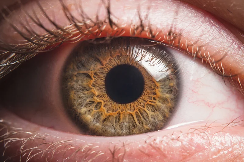
The cornea is a transparent outer tissue that covers the iris and the human eye. It consists of five layers and is covered with tears that protect it from microbes and provide it with food.

The cornea has two important functions:

 -First: protect the eyes from dust and microbes

-Second: It acts as a collecting surface for light rays until it falls on the lens of the eye and then gathers in one point on the retina to give a clear image.

What are the diseases that affect the cornea?

One of the most famous injuries that occur to the cornea and affect its efficiency is the presence of a foreign body in the eye or a laceration in the tissues and layers of the cornea as a result of exposure to get infected keratoconus is also one of the most common genetic diseases that affect the cornea and reduce visual acuity and is initially treated with contact lenses or Medical glasses and it can end up with a corneal graft to maintain the strength of vision in the eyes.

How does the injury occur?

The factors that lead to the occurrence of injury:

-chemical agents.

-yellowness.

-Diseases of the tissues of the eye, such as (Steven Johnson Syndrome).

-Chronic eye infections.

-Weakness in the fifth nerve, which is responsible for the sensation of the cornea.

-All of these factors eventually lead to the growth of blood vessels in the cornea and the loss of transparency of the cornea, and thus weakness severe in vision.

What is meant by corneal transplant?

Corneal transplant is one of the most successful ophthalmology surgeries, which reach 90%.The importance of corneal transplantation is to create a tissue that has transparency and thus maintain visual acuity.


Cases in which corneal transplantation is used

-All kinds of ulcers

-advanced keratoconus

-Injury or injury

-corneal clouds

What is meant by defects of visual acuity?

Visual impairment is divided into:

-Nearsightedness

-Farsightedness

-astigmatism

-Vision impairment due to aging

What is the Nearsightedness?

Research has shown that about 1/3 of the population suffers from Nearsightedness.

Reasons:

-The lack of light amplitude in one point in front of the retina due to the large size of the eye

-Symptoms: headache or occurrence around the eye, especially while driving a car or reading for a long time cure it Non-surgical treatment: glasses. Contact lenses

Surgical treatment: LASIK.

What is meant by long-sightedness?

Farsightedness occurs in about ¼ of the population

Causes: The light rays do not gather at one point on the retina, and the light rays gather at a point behind the retina, which causes blurring.

Symptoms: headache or occurrence around the eye cure it.

Wound treatment: using surgical laser.

Non-surgical treatment: glasses or contact lenses.

What is meant by astigmatism?

Symptoms: People with a small percentage may not notice it, but if it is not corrected

Severe astigmatism feels headache and pain in the eye with blurred vision

Treatment: Astigmatism can be corrected using an excimer laser, especially for the regular and mixed types

Regular astigmatism occurs when light rays fall on more than one point (location) on the retina

This causes the surface of the cornea to not rotate completely, which leads to blurring of vision and astigmatism

In cases of short and long-sightedness

It is divided into several types: regular – irregular – mixed

What is double vision associated with age?

Low vision associated with age is a natural symptom with increasing age, especially after the age of forty, and it can be corrected Wear glasses while reading.

What is Keratoconus?

The normal intraocular pressure causes the thin cornea to gradually bulge outward, which changes the shape of the spherical cornea.

This change of shape leads to a negative effect on the strength of vision and vision.

In some severe cases, this may lead to the inability to perform daily tasks such as reading, driving, etc.

Although this condition does not lead to complete blindness, it may lead to the inability to work in 20% of cases require corneal graft surgery to restore vision.

How does keratoconus affect vision?

In the early stages, a slight change in visual acuity occurs, after which the level of vision begins to fluctuate, and the patient needs to change his eyeglasses or contact lenses.

Apply repeatedly and at frequent intervals

The more outwardly bulging the cornea, the sharper the cone and the greater the amount of scattering of the light rays entering the eye, resulting in what is called astigmatism.

Regularly leads to visual impairment in a way that cannot be corrected with regular glasses

At this stage, it is necessary to use hard contact lenses. In the advanced stages, the use of glasses and hard lenses becomes tiring and useless.

A cloud, opacity, or ulcer forms on the surface of the cornea, and hydrocephalus of the cornea can occur, and then vision is greatly reduced.

In 20% of patients, corneal transplant surgery is required to treat advanced cases, and then another treatment method or some visual aids are required other to correct vision properly.

What are the symptoms of keratoconus?

-Blurring in the eye and changing the size of the glasses at frequent intervals

-Increased sensitivity to light

Difficulty driving at night

-Eye fatigue

-Headache and pain around the eyes

-Eye irritation and severe itching

How can diagnosing keratoconus?

-Consultation with an ophthalmologist.

-Complete eye examination.

-The measurement of the optical error determines its type and the degree of response to simple lenses.

-pachymetry is a measurement of the thickness of the cornea.

-Computerized corneal scan.


Looking forward

Let’s share with you our news and special offers of our services.


    Contact us


    Call us

    0-800-777-2331


    Visit us anytime

    27th Avenue, W2 3XE, New York


    Send us an email

    office@medicare.com


    Subscribe


    Sign up for Medicare newsletter to receive all the news offers and discounts from Medicare eye clinic.


      Social networks


      Facebook

      www.facebook.com/medicare


      Twitter

      #WeCare


      Instagram

      www.instagram.com/medicare


      Get in touch

      We are pleased to provide our distinguished services to you through several branches
      equipped with the best and latest medical technologies.


      Al-Dokki Branch


      Nasr-City Branch


      Beni-Suef Branch


      Contact us


      Call us

      0-800-777-2331


      Visit us anytime

      27th Avenue, W2 3XE, New York


      Send us an email

      office@medicare.com


      Subscribe


      Sign up for Medicare newsletter to receive all the news offers and discounts from Medicare eye clinic.


        Social networks


        Facebook

        www.facebook.com/medicare


        Twitter

        #WeCare


        Instagram

        www.instagram.com/medicare


        phone
        facebook